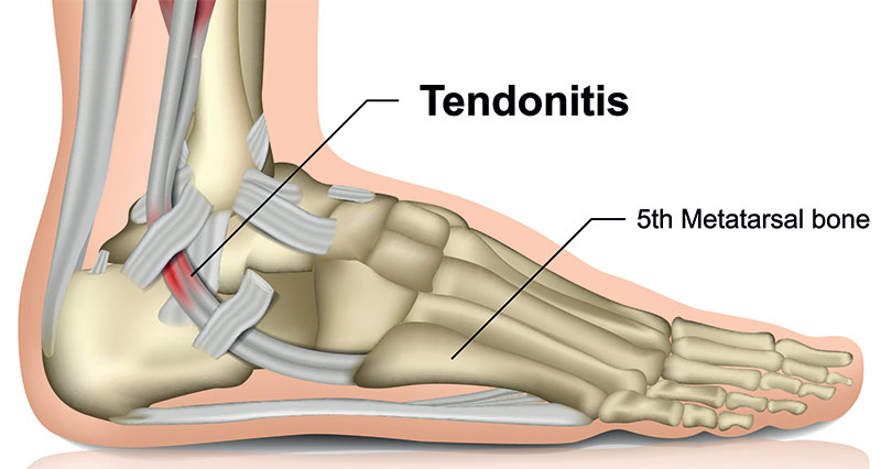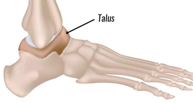
Osteochondral lesions of the talus are fractures of the cartilage on top of the Talus (ankle bone). Often occurring with a sprained ankle, it sometimes goes undiagnosed.
Signs and Symptoms
Symptoms of an Osteochondral lesion of the talus include
- Ankle pain
- Swelling
- Ankle locks or catches
- Joint stiffness
For an accurate diagnosis, you may need a bone scan, MRI or surgery.
What is an Osteochondral lesion of the talus?
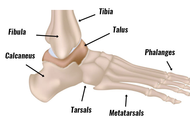
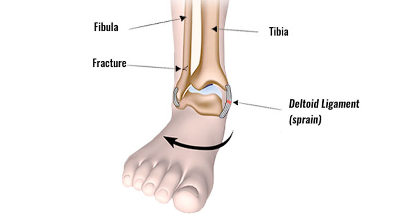
Osteochondral lesions or fractures of the cartilage which sits on top of the Talus (ankle bone), most commonly occur in combination with an ankle sprain. This is especially common if the injury occurs when landing on the ankle. The Tibia above compresses the top of the Talus, damaging the cartilage and protecting the bone.

Your physio or doctor may not initially diagnose Osteochondral lesions because it goes unnoticed with ankle sprain symptoms. It is only when your ankle improves to an extent but continues to be painful and swollen that they consider complications and investigate further.
Large fractures may be evident on X-rays. MRI scans, isotopic bone scans. However, CT scans will more likely pick up smaller lesions.
We grade Osteochondral lesions 1-5, with 1 being the least severe:
I – Subchondral fracture
II – Chondral fracture
IIa – Subchondral cyst
III – Chondral fracture with separate but not displaced fragments
IV – Chondral fracture with separate and displaced fragments
If your doctor does not detect an Osteochondral injury, it may eventually lead to Osteoarthritis of the ankle1.
Treatment of Osteochondral Lesions of the talus
Treat Grade I and II lesions conservatively (without surgery). In the past, your doctor might recommend immobilizing your ankle in a cast. However, a more modern approach is not to do this because mobilizing your joint (non-weight bearing) promotes cartilage healing.
Avoid weight-bearing activities. Instead, replace it with cycling or swimming to maintain fitness and keep your ankle mobile.
Grade I and II injuries that do not heal within 3 months may be put forward for surgery.
Grade III and IV injuries require an ankle arthroscopy (keyhole surgery) to remove the separated fragments.
When possible you should complete a full ankle rehabilitation program which includes strengthening, mobility and movement control exercises.
References & further reading
- Lee M, Kwon JW, Choi WJ, et al. Comparison of outcomes for osteochondral lesions of the talus with and without chronic lateral ankle instability. Foot Ankle Int 2015;36(9): 1050–7.